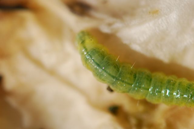
Remember the see-through green caterpillar from last week? I’ve been studying it some more.
After discovering the air passages or trachea were visible through the exoskeleton, I decided to look for other internal features.
 What about the dark green line in the middle of the back? (Photograph was taken looking down at the caterpillar.)
You might think that was the intestine, but I happen to know that the “heart” in insects is a long tube running through the dorsal surface (back). Could that be the “heart?”
Let’s take a look at this video taken from the perspective looking down at the caterpillar from the rear. Watch the green line.
If you look closely, you should be able to see the vessel expand and contract as hemolymph (the insect version of blood) is pumped through.
You might also notice that the pumping is not regular. It starts and stops.
The circulatory system in insects consists of a long tube called the dorsal vessel. The part running through the abdomen is designated as the heart.The aorta is located in the thorax and head.
Unlike ours, it is an open system. The hemolymph bathes the tissues inside the insect. When the heart pumps, the hemolymph moves into the dorsal vessel from the posterior or rear of the caterpillar and flows toward the head end.
In the dorsal vessel are openings known as ostia. Depending on position and type of insect, the ostia may be “incurrent” (hemolymph flows in) or “excurrent” (hemolymph flows out).
The hemolymph that exits goes back into the tissues, carrying in nutrients and moving away wastes.
It was pretty cool to see something I’d learned about in textbooks pumping away in real life.
Have you ever seen an insect heart in action?
Want to learn more? Try this article about the circulatory system in insects at North Carolina State.
